1
2
3
4
5
6
7
8
9
10
11
12
13
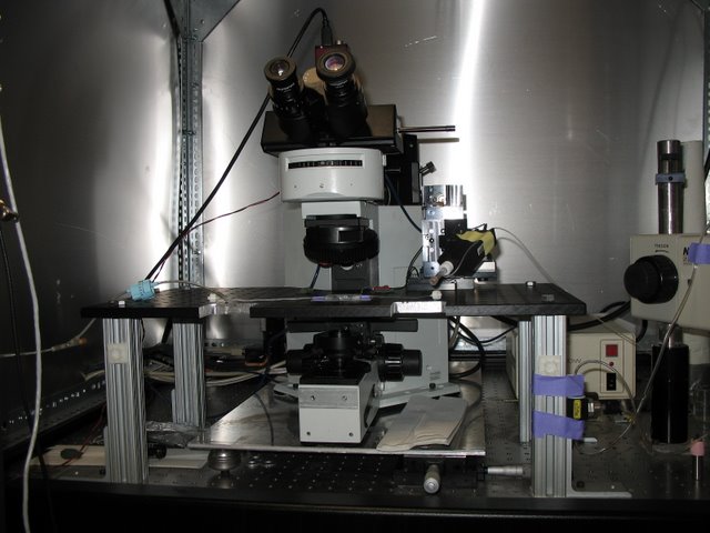
Meister Lab: This Olympus microscope has been modified to allow for patch clamp recordings from retinal ganglion cells. The stage has been replaced with a stable fixed table onto which the preparation and manipulator are mounted. Translating the F.O.V. is accomplished by moving the microscope.
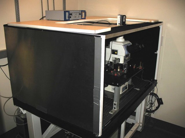
Meister Lab: The same microscope from behind. The trans-illumination lamp has been removed and replaced with a digital projector. This allows patterned images to be presented to the retain while cellular responses are recorded. Incompatible E.M.F. sources have been removed or modified.
Lichtman Lab: Similar modifications were made to this Nikon microscope to allow for patch and extracellular recordings. Custom Faraday cage, fixed stage, and microscope translator are visible.
Lichtman Lab: Custom objective changing/focusing unit was added to account for the Nikon’s lack of a turret focus.
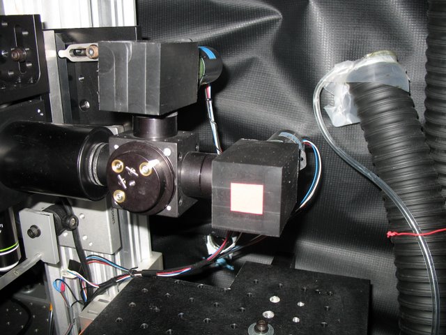
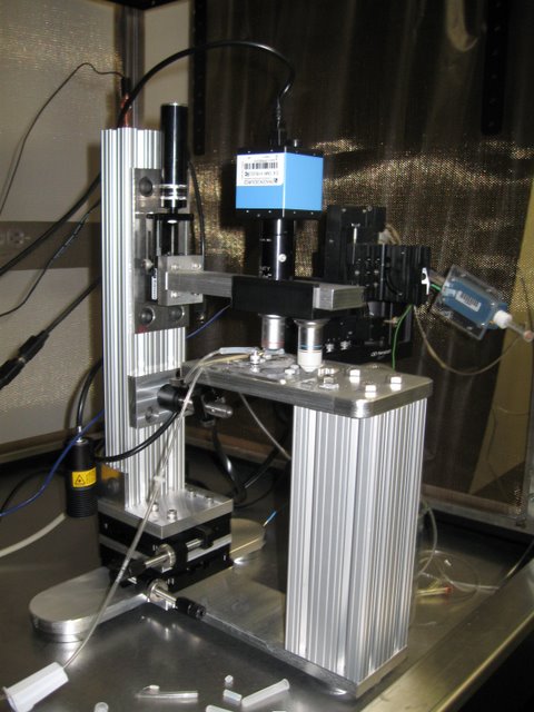
Murthy Lab: We have produce numerous devices for the many custom built 2 photon microscopes that exist in CBS. Here two PMT mounts are shown as part of a two channel, two photon microscope.
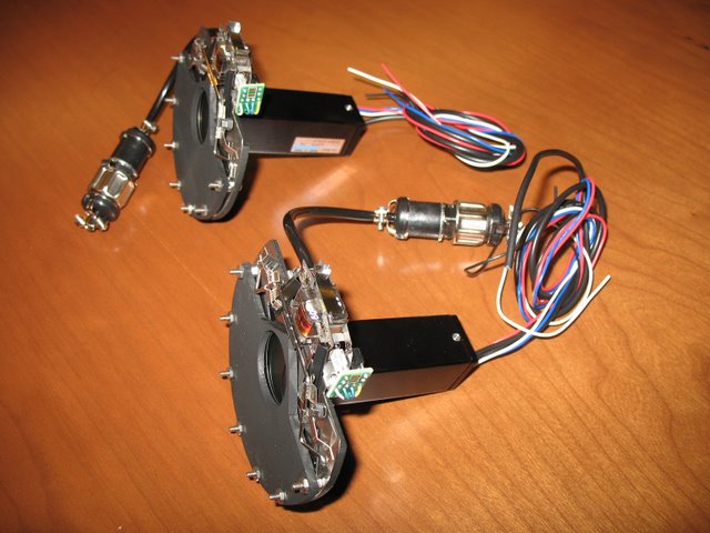
Reid, Wilson, Engert Labs: GaAs detectors used in 2 photon microscopy are very sensitive and easily damaged by excess light. These custom shutters are part of a protection system. They are designed to be very thin so as not to reduce the amount of light arriving at the detector. The central aperture is 1” in diameter.
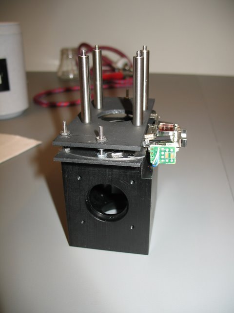
Wilson Lab: A variation of the shutter design, integrated into a beam splitting cube.
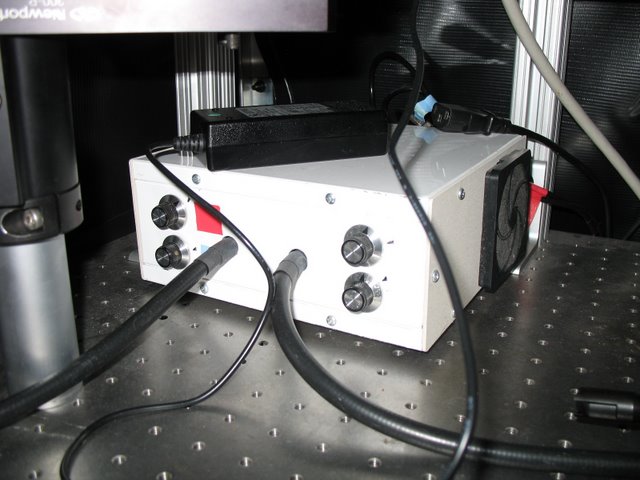
Meister/Murthy Labs: A dual fiber, two channel L.E.D. light source capable of rapidly switching between N.I.R. and 465 nm illumination. By synchronizing illumination with video acquisition it was possible to simultaneously measure olfactory neural responses via intrinsic and synaptophluorin signals.
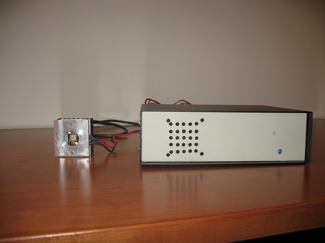
Murthy Lab: A mount and driver electronics for a 5W blue Phlat light L.E.D. manufactured by Luminus. Lower power devices have been constructed for the Uchida, Murthy, Engert, and Meister labs.
Meister Lab: A custom designed and fabricated photolithography station. This tool allows the Meister lab to fabricate their 61 channel planar electrodes arrays
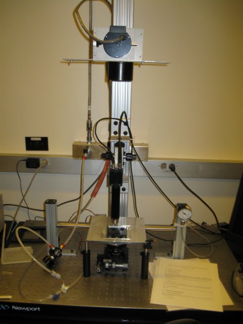
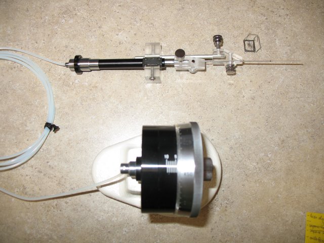
Murthy/Uchida Labs: Adaptors to convert an extracellular electrode manipulator into a nanoliter injection system used for virus delivery.
Landisman Lab: A slice patch clamp rig with integrated microscope. This microscope was designed to deliver D.I.C.-like image quality for visually guided patch recording. It provides a very stable and open platform and cost one-sixth the price of a traditional scope.
Landisman Lab: Living neurons imaged using our patch microscope. Expensive D.I.C. optics are replaced with shallow angle N.I.R. illumination.